What Are Nucleic Acids and Why Do They Matter?
Nucleic acids, like carbohydrates, proteins, and lipids, are essential macromolecules — large molecules that play crucial roles in the structure and function of all living things. The four major macromolecules are:
Nucleic acids (you're here!)
The other three macromolecules build parts of the organism and run reactions. Nucleic acids do something different: they carry information. Every instruction needed to build and operate a living organism is stored in the order of a few simple subunits strung together in a line. Change that order and you can change the organism.
In fact, many biologists think information came first. The RNA world hypothesis proposes that life began with nucleic acids, specifically RNA, before proteins or DNA existed, because RNA can both store information and speed up chemical reactions. We'll revisit that at the end.
What Are Nucleic Acids Made Of?
Unlike lipids, nucleic acids follow the monomer rule cleanly. The monomer of nucleic acids is called the nucleotide. If you link many nucleotides together, you get a polymer.
Every nucleotide has exactly three parts:
A phosphate group
A five-carbon sugar
A nitrogenous base
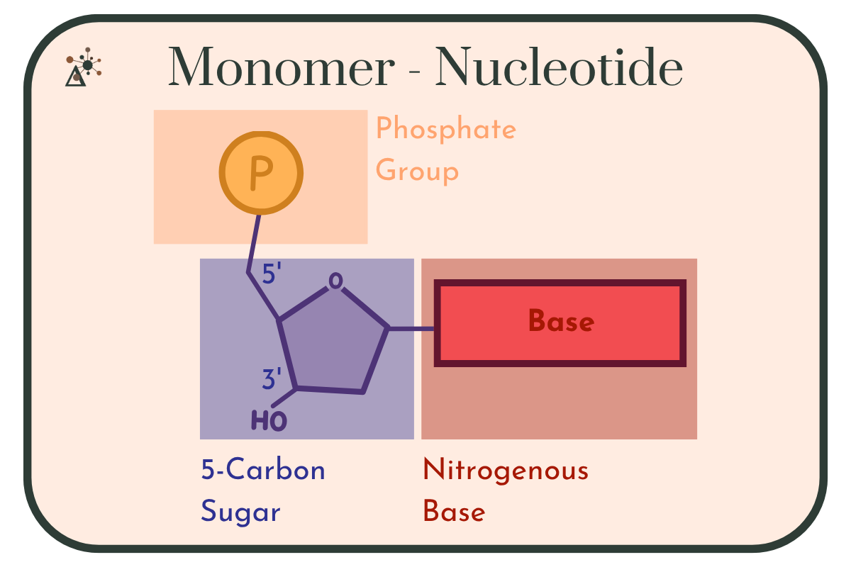
Image 1: Diagram of a single nucleotide showing its three components in color-coded blocks: an orange phosphate group, a purple five-carbon sugar drawn as a pentagon with its 5-prime and 3-prime carbons labeled and a hydroxyl group at the 3-prime position, and a red rectangle representing the nitrogenous base. The phosphate attaches to the 5-prime carbon and the base attaches on the opposite side of the sugar.
Cells make a wide variety of nucleotides, but in an introductory biology course the focus stays on two: ribonucleotides and deoxyribonucleotides. The difference between them comes down to a single oxygen atom.
Two Sugars, Two Nucleic Acids
The sugar in a ribonucleotide is ribose. The sugar in a deoxyribonucleotide is deoxyribose. The prefix deoxy- means "lacking oxygen," and it differentiates the two sugars: deoxyribose is missing one oxygen at the 2' carbon, where ribose carries a hydroxyl (–OH) group.
That one missing oxygen is the difference between the two nucleic acids:
Ribose → RNA (ribonucleic acid)
Deoxyribose → DNA (deoxyribonucleic acid)
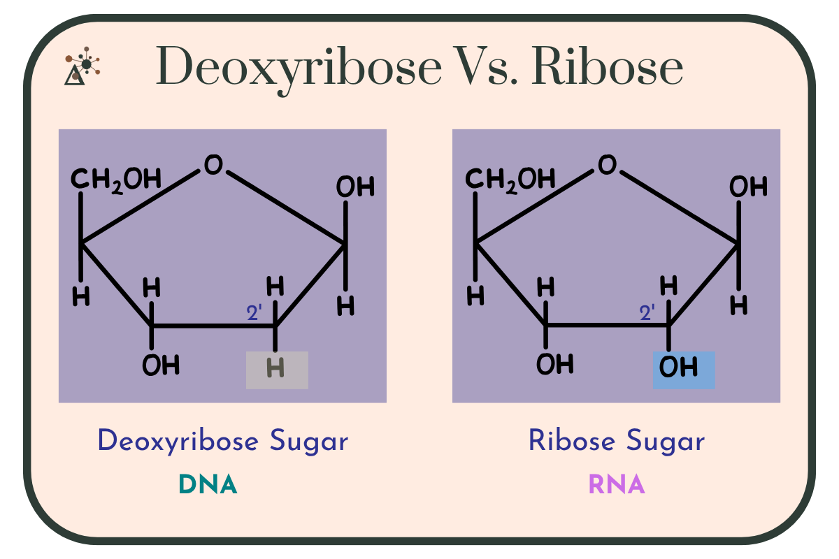
Image 2: Two five-carbon sugar structures side by side, drawn identically except at one position. Deoxyribose, labeled DNA, has a hydrogen atom highlighted in grey at the 2-prime carbon. Ribose, labeled RNA, has a hydroxyl group highlighted in blue at the same 2-prime carbon. The 2-prime carbon is labeled on both.
The Nitrogenous Bases
The third component, the nitrogenous base, is what actually carries the information. Bases fall into two structural groups:
Purines have a double-ring structure. There are two: adenine (A) and guanine (G).
Pyrimidines have a single-ring structure. There are three: cytosine (C), thymine (T), and uracil (U).
Adenine, guanine, and cytosine appear in both DNA and RNA. Thymine appears only in DNA, and uracil appears only in RNA, filling the same role thymine does.
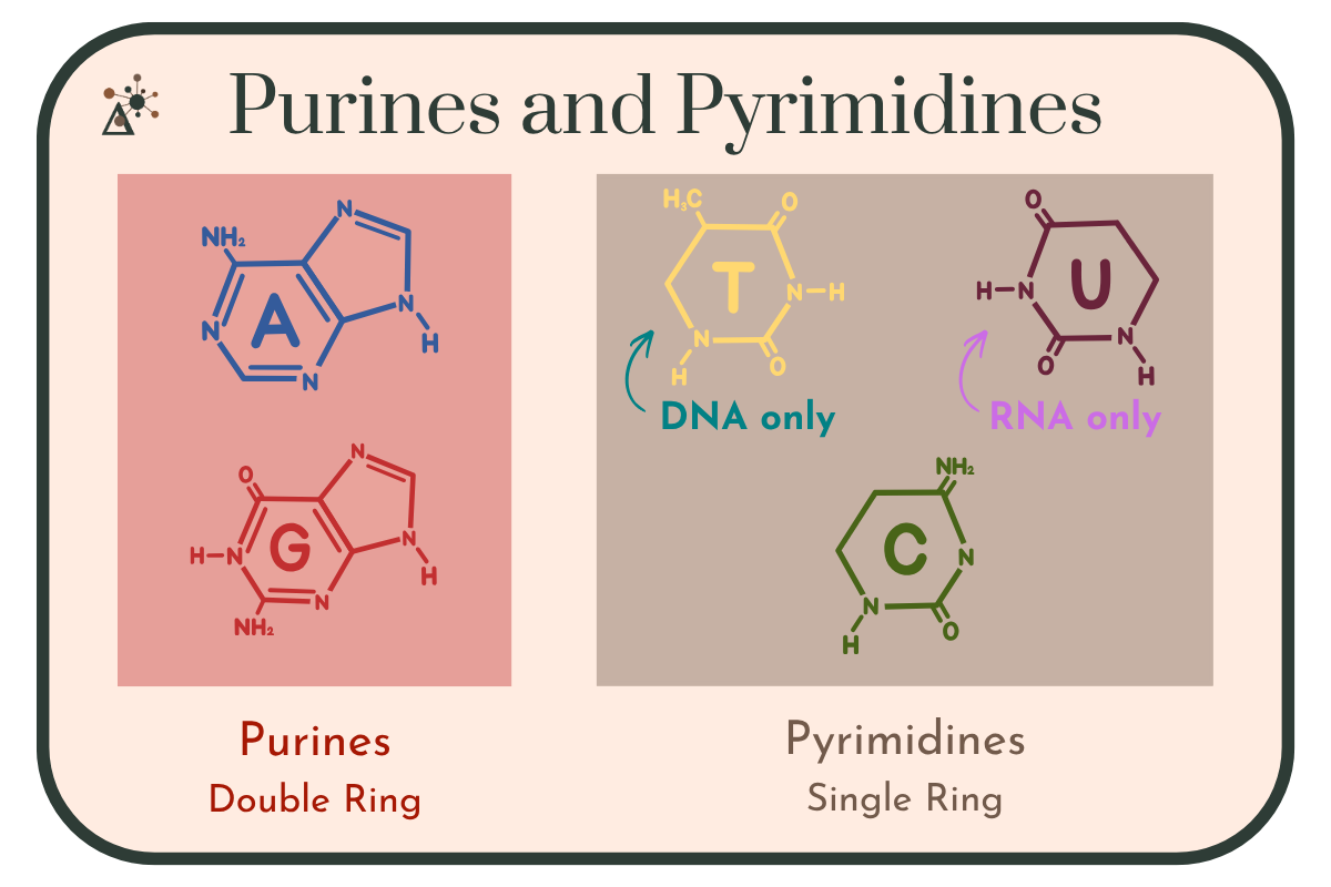
Image 3: Two panels comparing the nitrogenous bases. The left panel, labeled Purines, Double Ring, shows adenine and guanine, each drawn as two fused rings. The right panel, labeled Pyrimidines, Single Ring, shows thymine, uracil, and cytosine, each drawn as a single ring. Thymine is tagged DNA only and uracil is tagged RNA only.
Quick check: It is common to think that pyrimidines are the double-ring structure because pyrimidine is the longer word. One way to keep it straight: CUT the PY — Cytosine, Uracil, and Thymine are the pyrimidines. Everything left over is a purine.
Linking Nucleotides: The Sugar-Phosphate Backbone
Nucleotides join together through a condensation reaction (also called a dehydration reaction), the same type of reaction that builds the other macromolecule polymers. The bond forms between the phosphate group of one nucleotide and the sugar of the next, specifically at the 3' carbon.
The resulting bond is called a phosphodiester linkage.
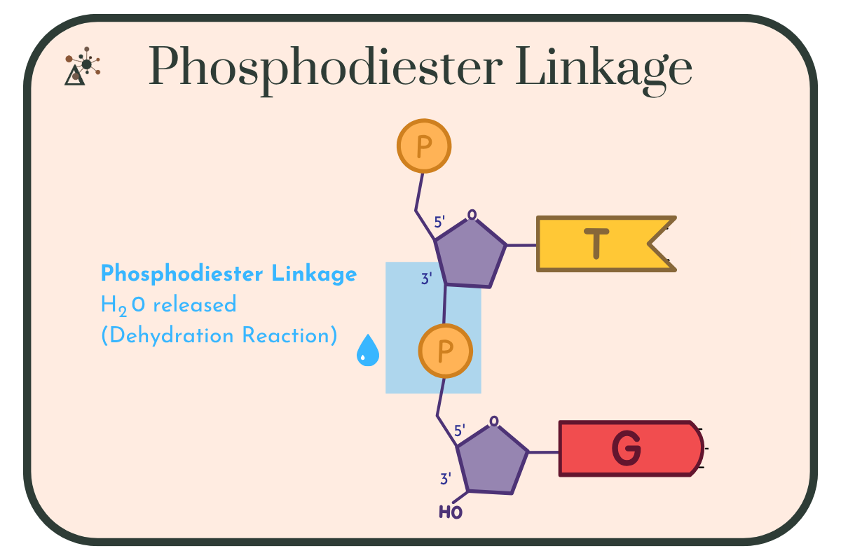
Image 4: Diagram of two nucleotides joining together. The upper nucleotide carries a phosphate at its 5-prime carbon and a thymine base; its 3-prime oxygen bonds downward to the phosphate of the lower nucleotide, which carries a guanine base and a free hydroxyl at its 3-prime carbon. The bond region is highlighted, with a water droplet indicating that water is released in this dehydration reaction.
Because every linkage connects a phosphate to a sugar, a long chain of nucleotides has an alternating sugar–phosphate–sugar–phosphate pattern running down its length. This is the sugar-phosphate backbone, and the bases hang off it like teeth on a comb.
The Backbone Has a Direction
There is a feature present in nucleic acids that has no equivalent in lipids or carbohydrates: a nucleic acid strand has a direction.
One end of the strand has a free phosphate group attached to the 5' carbon. The other end has a free hydroxyl group on the 3' carbon. These are called the 5' end and the 3' end, and they are not interchangeable.
By convention, sequences are always written in the 5' → 3' direction. When you see a sequence written as ATGCC, the A is at the 5' end.
The image below shows the directionality of DNA. As we’ll explore further in the article, DNA is both double stranded and antiparallel, meaning that the strands run in opposite directions. One strand runs in the 5' → 3' direction, while the second strand in the 3' → 5' direction.
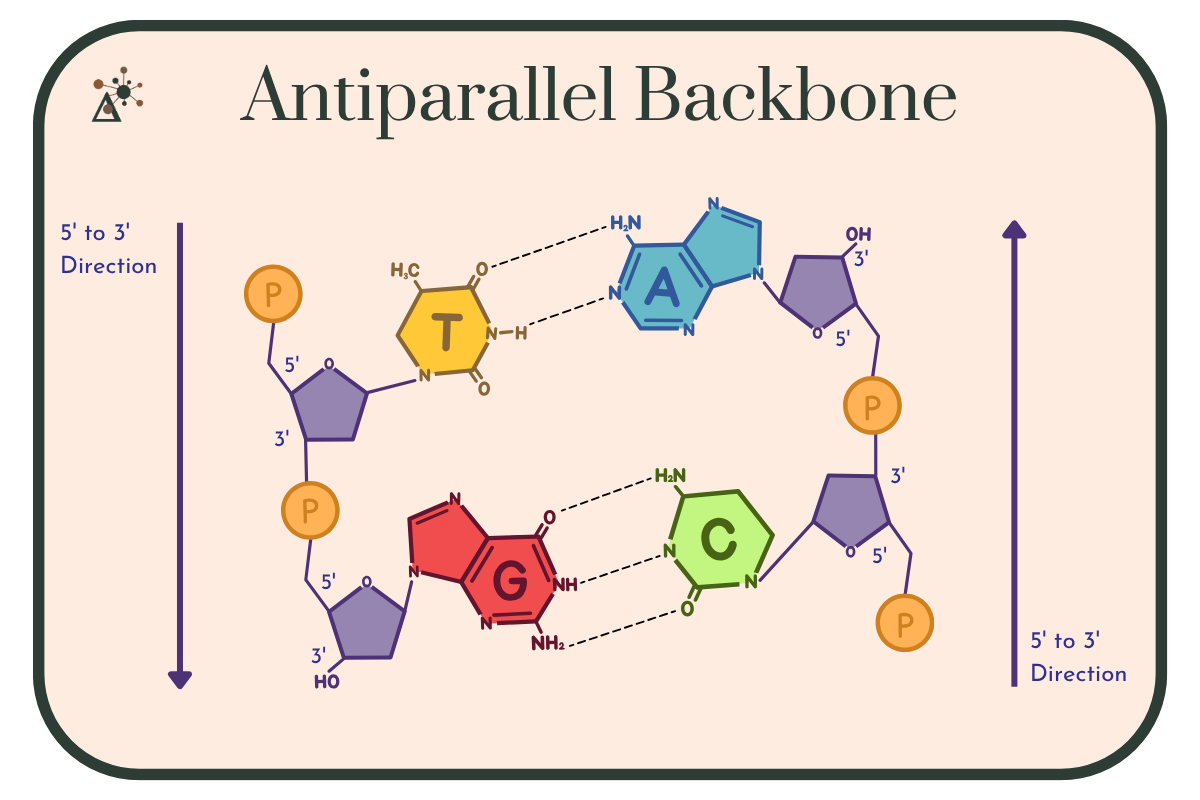
Image 5: Two DNA strands running in opposite directions, each marked with an arrow showing its 5-prime to 3-prime direction: the left strand runs downward and the right strand runs upward. Orange phosphates alternate with purple sugars along both backbones. Thymine pairs with adenine across two dashed hydrogen bonds, and guanine pairs with cytosine across three.
The Major Groups of Nucleic Acids
Both DNA and RNA have a primary structure. It is the sequence of nucleotides in the strand. Both also have a secondary structure, formed by hydrogen bonding between nitrogenous bases. But they use that secondary structure very differently.
DNA
In 1953, James Watson and Francis Crick published a model of DNA's structure that explained, in one stroke, how genetic information could be stored and copied.
They did not do it alone. Their model depended on X-ray diffraction data produced at King's College London by Rosalind Franklin and her PhD student Raymond Gosling, and on work by Maurice Wilkins. It also depended on Erwin Chargaff's finding that in any sample of DNA, the amount of adenine equals the amount of thymine, and the amount of guanine equals the amount of cytosine, now known as Chargaff's rules.
Franklin and Gosling worked by drawing out a fine fiber of DNA and firing X-rays through it, then recording how the beams scattered. Their best-known result is the image known as Photograph 51.
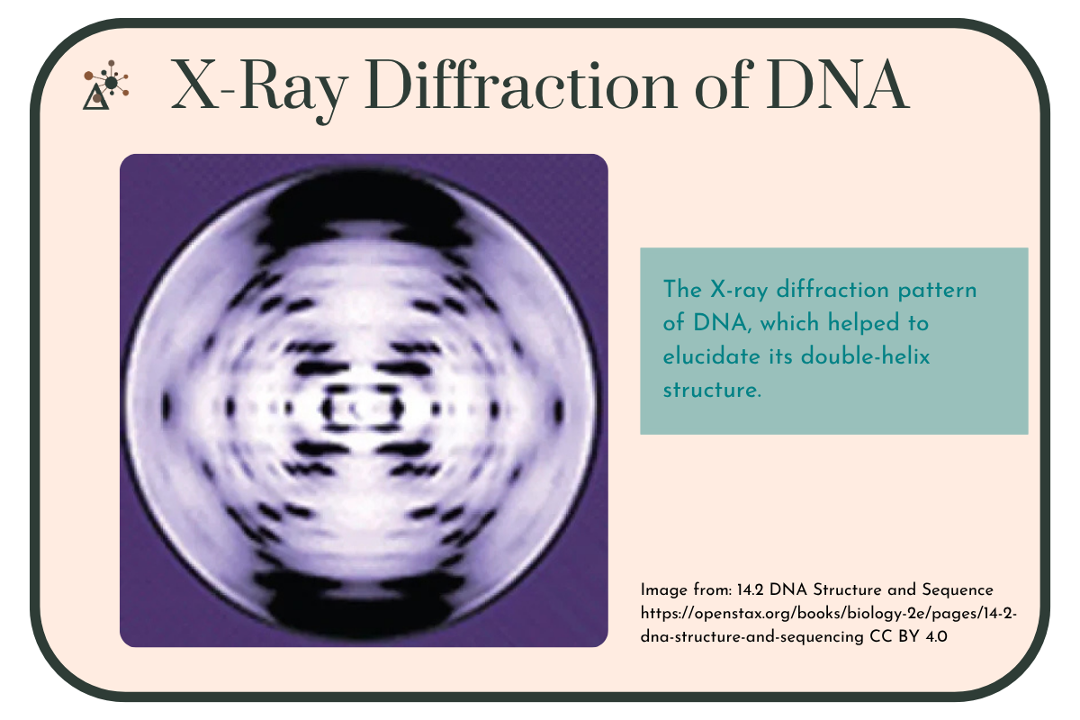
Image 6: An X-ray diffraction photograph of DNA, showing a roughly circular pattern of dark and light spots with heavy dark bands at the top and bottom. Patterns like this one allowed researchers to work out the double-helix structure.
The image above is not Photograph 51. That famous photograph — taken by Raymond Gosling in Franklin's lab — is a blurrier image of a different DNA form, and it's still under copyright, which is why you won't find it reproduced in most free educational materials. It's also, according to the historians who re-examined the record, probably not the decisive piece of evidence the popular story makes it out to be.
You can find the image here: King’s College London
Note on History: You may have heard that Watson and Crick stole Franklin's data and that she failed to understand her own results. That version has been challenged. In a 2023 opinion piece in Nature, Matthew Cobb and Nathaniel Comfort argue that Franklin should be remembered not as the victim of the double helix but as an equal contributor to solving its structure. Drawing on a colleague's letter and a previously undescribed draft article written for Time magazine, they make the case that she knew her data had been shared with the Cambridge group and was working as a peer rather than an unwitting victim.
Not every historian agrees. Others maintain that Watson and Crick's use of her unpublished work was improper, and that she was denied recognition partly because she was a Jewish woman. What almost everyone agrees on is the credit itself: Franklin's data was essential to the discovery, and she did not receive appropriate public recognition for it in her lifetime. When the Nobel Prize was awarded in 1962 to Watson, Crick, and Wilkins, Franklin had already died of ovarian cancer in 1958 at age 37, and Nobel Prizes are not awarded posthumously.
The Double Helix
The model showed DNA as two strands wound around each other in a double helix, with these key features:
The two strands are antiparallel. One runs 5' → 3' while the other runs 3' → 5'.
The sugar-phosphate backbones face outward, toward the surrounding water.
The bases face inward, stacked in the interior of the helix.
Bases pair specifically: a purine always pairs with a pyrimidine. A pairs with T, and G pairs with C. These are called complementary base pairs.
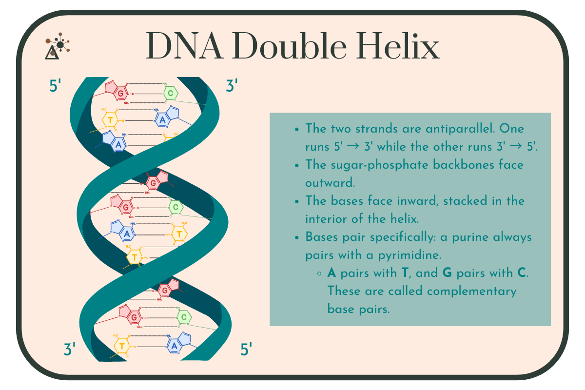
Image 7: A right-handed DNA double helix drawn as two teal ribbons winding around each other, with color-coded base pairs joined by dashed hydrogen bonds in the interior. The strands are labeled 5-prime and 3-prime at opposite corners, showing that they run antiparallel, and the sugar-phosphate backbones face outward while the bases face inward.
A purine (double ring) paired with a pyrimidine (single ring) is always three rings wide, so the helix stays a constant width down its whole length. Pairing two purines would bulge, and pairing two pyrimidines would pinch.
The pairs are held together by hydrogen bonds — two between A and T, three between G and C. Because of the extra bond, G-C pairs are harder to pull apart, so DNA regions rich in G and C take more energy to separate.
Quick check: Hydrogen bonding gets most of the attention, but it isn't the only thing holding the helix together. The stacked bases in the interior also cling to each other through base-stacking interactions, and these contribute at least as much to the stability of the double helix as the hydrogen bonds do. The hydrogen bonds provide the specificity, who pairs with whom. The stacking provides much of the stability.
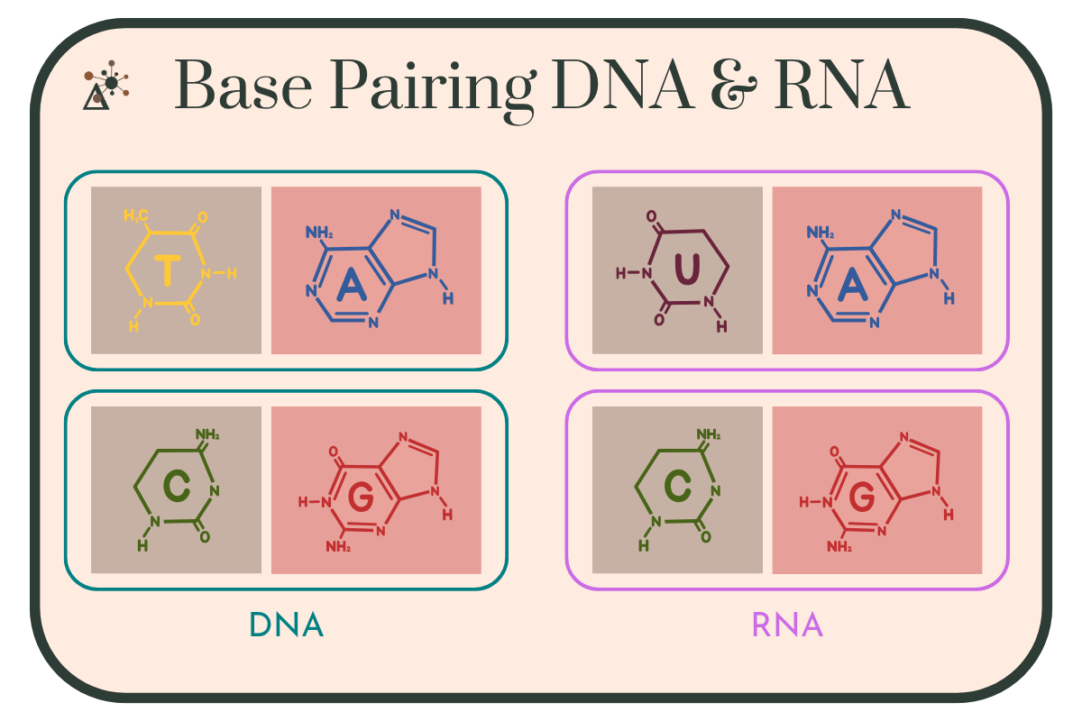
Image 8: Four base pairs grouped into two sets. The DNA set, outlined in teal, shows thymine paired with adenine and cytosine paired with guanine. The RNA set, outlined in magenta, shows uracil paired with adenine and cytosine paired with guanine. Uracil takes the place of thymine in RNA.
Hydrophobic Inside, Hydrophilic Outside
Nitrogenous bases are relatively nonpolar, while the sugar-phosphate backbone is charged and highly polar. Tucking the bases into the interior and turning the backbone outward puts each part of the molecule where it's chemically comfortable.
The result is a molecule with a hydrophobic interior and a hydrophilic exterior, which means DNA as a whole dissolves readily in the watery interior of a cell.
If that arrangement sounds familiar, it should. It's the same logic that drives phospholipids into a bilayer: nonpolar regions hide from water, while the polar regions face it. Two completely different macromolecules, same underlying principle.
RNA
RNA is built the same way as DNA, from nucleotides linked by phosphodiester bonds, with three differences: ribose instead of deoxyribose, uracil instead of thymine, and most importantly RNA is typically single-stranded.
That single strand is free to fold back on itself, and RNA is described as having up to four levels of structure:
Primary structure: the sequence of nucleotides along the sugar-phosphate backbone.
Secondary structure: shapes formed when complementary bases on the same strand pair with each other. The most common is the hairpin (or stem-loop), where a strand doubles back and pairs with itself.
Tertiary structure: the full three-dimensional fold of the molecule.
Quaternary structure: associations between multiple separate RNA molecules.
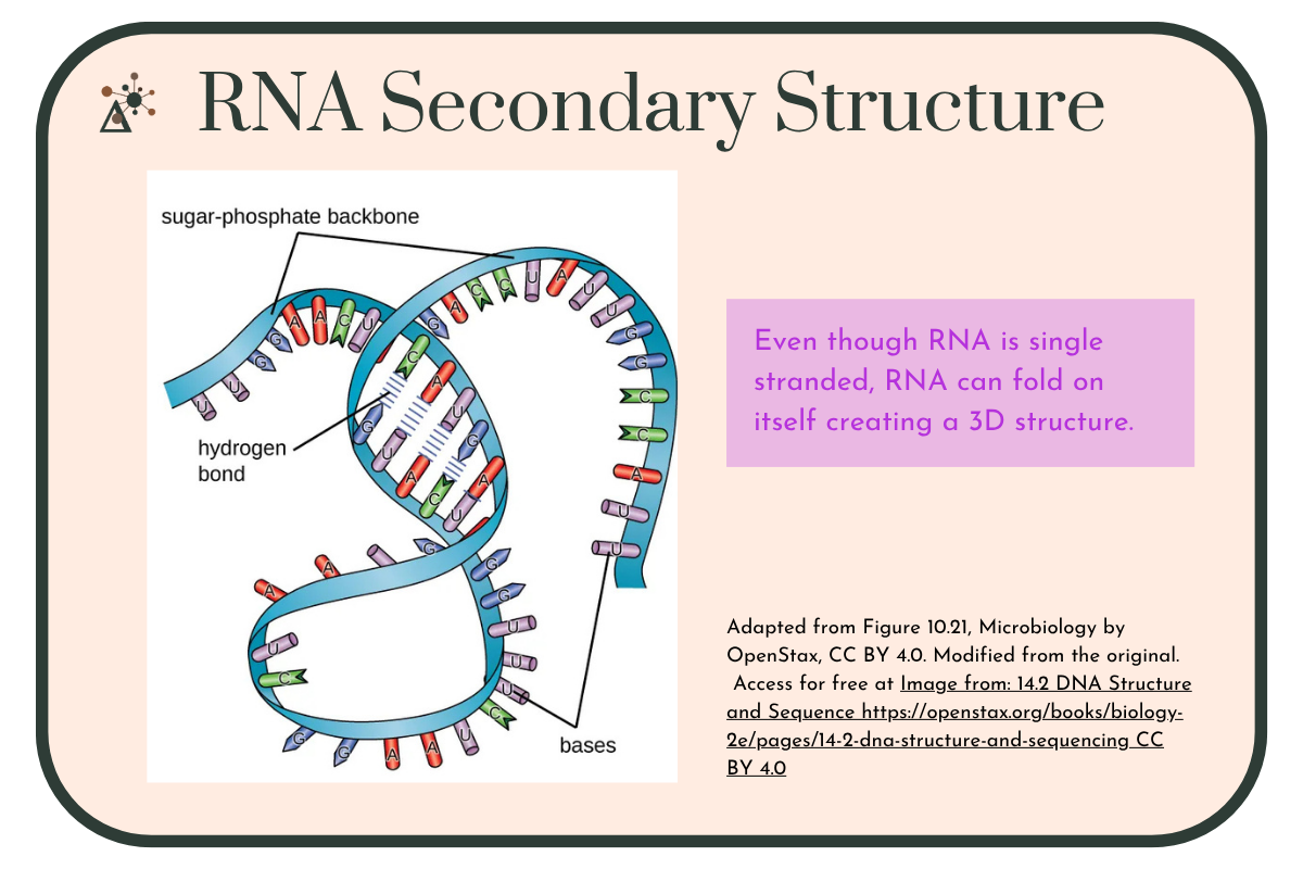
Image 9: A single strand of RNA folded back on itself, with its sugar-phosphate backbone drawn as a ribbon and its bases labeled A, U, G, and C. Short regions where complementary bases meet are held together by hydrogen bonds, shown as small parallel lines, holding the folded shape in place.
This structure is similar to the protein structure. But RNA's structural range is different: with four bases to work with instead of twenty amino acids, RNA folds are not as diverse or as complex as protein folds.
What Do Nucleic Acids Do?
As with every macromolecule, structure dictates function.
DNA: Stores and Transmits Information
DNA's job is to hold information stably and pass it on accurately.
Storage: Information is encoded in the sequence of nucleotides. With four bases, a stretch of DNA just ten nucleotides long has over a million possible sequences. A human genome is roughly 3 billion base pairs.
In every cell examined to date, from the smallest bacteria to the largest redwoods, DNA carries the information required for the organism's growth and reproduction.
Transmission: Because A only pairs with T and G only pairs with C, each strand of a double helix specifies the sequence of the other. One strand acts as a template, and a matching complementary strand can be built against it. This is how a cell copies its DNA before dividing, and it was the single most powerful implication of the 1953 model: the structure explains the copying mechanism.
Stability: DNA is chemically stable (more so than RNA, thanks to that missing 2' hydroxyl), which makes it well suited to long-term information storage.
Quick check: "Stable" doesn't mean "damage-proof." Every cell in your body sustains DNA damage every day from ordinary metabolism, radiation, and chemical exposure. What keeps genomes intact isn't chemical toughness alone but an extensive set of DNA repair enzymes working continuously. And because the two strands are complementary, damage to one strand can be repaired using the other as a reference. Stability is partly a property of the molecule and partly an active, ongoing process.
RNA: Does the Work
If DNA is the archive, RNA is everything else. RNA performs key roles in information processing.There are several important types of RNA:
Messenger RNA (mRNA) carries a copy of a gene's sequence out of the nucleus to be read.
Transfer RNA (tRNA) brings the correct amino acid to the ribosome during protein synthesis.
Ribosomal RNA (rRNA) forms the structural and catalytic core of the ribosome. The ribosome is a ribozyme: the chemical step that joins amino acids together is catalyzed by RNA, not by protein.
Regulatory RNAs control which genes get expressed and how much protein is made.
Defensive RNAs help cells recognize and destroy viral genetic material. Bacterial CRISPR systems use short RNA guides to identify and cut invading viral DNA. CRISPR is the same mechanism now adapted into the gene-editing tools used in labs and clinics worldwide.
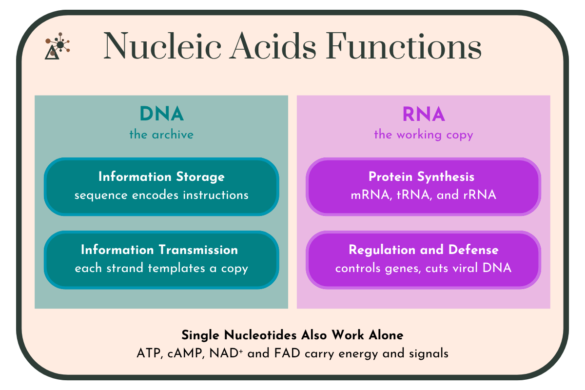
Image 10: Two-panel diagram comparing nucleic acid functions. The DNA panel, labeled "the archive," lists information storage, in which the sequence encodes instructions, and information transmission, in which each strand serves as a template for a copy. The RNA panel, labeled "the working copy," lists protein synthesis carried out by mRNA, tRNA, and rRNA, and regulation and defense, in which RNA controls gene expression and targets viral genetic material. A panel below notes that single nucleotides also work alone, with ATP, cAMP, NAD⁺, and FAD carrying energy and signals.
Nucleotides Also Work Alone
Not every nucleotide ends up in a polymer. Individual nucleotides and their relatives do essential jobs on their own:
ATP is the cell's primary energy currency.
cAMP carries signals inside cells.
NAD⁺ and FAD shuttle electrons through cellular respiration.
So when you meet ATP when learning about cellular respiration, you're meeting a nucleotide.
Why Nucleic Acids Matter
From one repeating subunit, a nucleotide, cells get:
Information storage in the sequence of DNA
Inheritance through complementary base pairing and templated copying
Protein synthesis carried out by mRNA, tRNA, and rRNA
Gene regulation and cellular defense through regulatory and guide RNAs
Energy and signaling from individual nucleotides like ATP and cAMP
That first one deserves emphasis. Proteins do the work of a cell and lipids draw its boundary, but neither can be built without instructions, and neither can pass those instructions to the next generation. Nucleic acids are how life remembers.
It's also why the RNA world hypothesis is so appealing. RNA can store information and catalyze reactions, the ribosome is living proof, which means RNA could in principle have done both jobs before DNA and proteins specialized. Researchers continue to test and refine that idea, and many current models propose that RNA and short peptides emerged together rather than RNA acting entirely alone. It's an open question, which is part of what makes it worth asking.
Understanding nucleic acids is also important in the field of medicine. Reading DNA sequences lets us diagnose genetic disease. CRISPR, borrowed from a bacterial RNA defense system, lets us edit genes directly. mRNA vaccines work by delivering a temporary strand of RNA that instructs your cells to build a viral protein for your immune system to learn from. Every one of those technologies started as a basic question about what these molecules are and how they're put together.
Nucleic acids are one of the four major macromolecules — alongside proteins, carbohydrates, and lipids — and they're the reason there is such a thing as inheritance at all.
CreditsOpenStax. (2021). Biology 2e, Section 14.2 DNA Structure and Sequencing. https://openstax.org/books/biology-2e/pages/14-2-dna-structure-and-sequencing. Licensed under CC BY 4.0 (https://creativecommons.org/licenses/by/4.0/). Image reproduced without modification. OpenStax. (2025). Microbiology, Section 10.3 Structure and Function of RNA, Figure 10.21. https://openstax.org/books/microbiology/pages/10-3-structure-and- function-of-rna. Licensed under CC BY 4.0 (https://creativecommons.org/licenses/ by/4.0/). Figure adapted: [cropped and background removed]. Access for free at https://openstax.org/books/microbiology/pages/1-introduction OpenStax. (2021). Biology 2e, Section 3.5 Nucleic Acids. https://openstax.org/books/biology-2e/pages/3-5-nucleic-acids. Licensed under CC BY 4.0 (https://creativecommons.org/licenses/by/4.0/). Cobb, M., & Comfort, N. (2023). What Rosalind Franklin truly contributed to the discovery of DNA's structure. Nature, 616, 657–660. https://www.nature.com/articles/d41586-023-01313-5Sullivan, W. (2023, April 28). What we're still learning about Rosalind Franklin's unheralded brilliance. Smithsonian Magazine. https://www.smithsonianmag.com/smart-news/what-were-still-learning-about- rosalind-franklins-unheralded-brilliance-180982058/